When assessing pathologies present in cells, such as cancer or various tumors, nuclear medicine is much more accurate. However, patients who must undergo such diagnostic tests think that nuclear appeal can be dangerous and radioactive for them and their environment.
To bring some light on nuclear medicine, we will explain what is and what PET and PET-CT scans consist of, which are the most common in oncology, cardiology, and neurology and the most unknown.
What is a PET Scan?
A CT scan of positron emissions, or PET, is one of the types of CT scans that are used today to get an accurate picture of the patient’s body inside. In this case, a small amount of radioactive substance is used. Despite being part of those known as CT, PET is not focused on bone anatomy or soft parts of the body but on the metabolic activity of the cells under study. The target? Diagnosing patterns that show the presence of tumors in cells by obtaining a three-dimensional image.
Thus, through the intravenous inclusion of this small amount of radioactive, both healthy cells and possible cancer cells, react, and do so differently. Hence the importance of the reactive subject and the use of this type of non-invasive diagnostic test. Thanks to this type of testing, oncologists can follow up more closely and more accurately on the evolution of their patients.
A PET can determine blood flow, oxygen consumption, and even the presence of blood sugar, so it is not only used for oncology but also for the study of cardiovascular pathologies and even for neurology.
The most interesting thing about this type of nuclear medicine test is the possibility of detecting all these anomalies in the early stages of the disease, thus gaining time, especially from cancer and possible cases of heart attacks and thrombi. In the three pathologies, time is key to shortening them by minimizing side effects on patients.
What a PET-CT scan consists of
Sometimes, the doctor chooses to jointly perform a PET test and the CT, since the same type of diagnostic device is used in both, even if the procedure is slightly different. Thus, the PET-TAC is two diagnostic tests in one, where you first get the images of the PET scan and then the CT scans. The receiving computer is responsible for unifying the images obtained from each of the tests, creating a three-dimensional image.
In addition to being determinant in oncology cases, it is one of the most commonly used diagnostic tests in neuropsychiatric diseases (Alzheimer’s, Parkinson’s, Epilepsy).
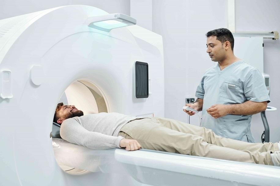
To get tested, the patient lies on the stretcher with an area to study inside the ring of the device. The patient has nothing to do but is still there because it is the stretcher that moves at all times for the taking of the images. In this case, the CT scan is first and then the PET.
When a PET exam is done?
That the doctor sends you to do a PET test does not imply that you have seen any suspicious element of being carcinogenic that you have not been told. Although it is true that this type of evidence, due to its nature and result, is more common in oncology consultations.
More specifically, the PET test helps the specialist both in the detection of cancer – many times even in the early stages of the disease – as it has verified the scope and dissemination (ramifications or metastasis), follows up on treatment, and confirms reproduction after elimination.
On the other hand, cardiology and neurology also often use this type of nuclear medicine test. In the case of cardiologists, to see the effects of a myocardial infarction on the heart muscle and to assess whether there are recoverable areas in a revascularization treatment. As for the neurologist, you can use this test to evaluate how the brain is working with memory problems, epilepsy, or similar neurological pathologies.
How a PET is made?
To carry out the PET test, the patient, as in the PET-TAC, lies on the stretcher, almost always facing up unless the doctor specifies this in the request or test steering wheel. It is the stretcher that moves through the hole of the CT shoe, so the patient’s only role is to remain very still for the duration of the test, which does not usually exceed 30 minutes.
Since it is done in an open device, it does not usually give a feeling of claustrophobia to patients. Nor are the continuous and annoying noises heard in an MRI.
However, here you also have to take off your clothes and put on the robe offered by the auxiliaries. Of course, you can’t introduce any metal objects or implants. Even the dentures can’t be carried at the time of the test.
In the case of PET, “the activator” is given 60 minutes before testing, either orally, intravenously, or through an inhaler.
Once the test is passed, the technician can ask the patient for extra time to check that the shot is valid and can work with it. Otherwise, the patient could be asked to repeat it.
When will I know the test results?
As with the CT tests, the images taken by the device can be seen immediately. However, to issue the diagnosis, the physician must study them and cross-examine them with the tests and other tests he may have requested. Your doctor will tell you or you will be referred to the consultation to know the results by medical citation.
Disclaimers: This information does not in any case replace the diagnosis or prescription by a doctor. It is important to go to a specialist when symptoms occur in case of illness and never self-medicate.

