I’m sure on more than one occasion you’ve heard from a friend or relative, and even yourself, that an MRI must be performed. You know what it’s about? Is it the same as MRI? The truth is that this non-invasive diagnostic examination has become one of the discoveries of the 20th century.
This diagnostic technique is very useful to obtain high precision images of the inside of the body without barely causing discomfort to the patient. Here we tell you what an MRI is and how it is done.
What is an MRI?
An MRI is a non-invasive diagnostic test that allows specialists to detect the presence of diseases and pathologies in the body. Thanks to the MRI, the physician has an accurate picture of the body structures.
Unlike CT scans, an MRI does not use X-rays to get the image inside the body, making it a safer diagnostic test. In fact, thanks to the MRI machine you can blur dozens of images of the area under study, so the accuracy of diagnosis increases.
Since it was invented in 1971 – its creators were therefore awarded the Nobel Prize in Medicine – it has become one of the safest and most accurate diagnostic techniques of medical science.
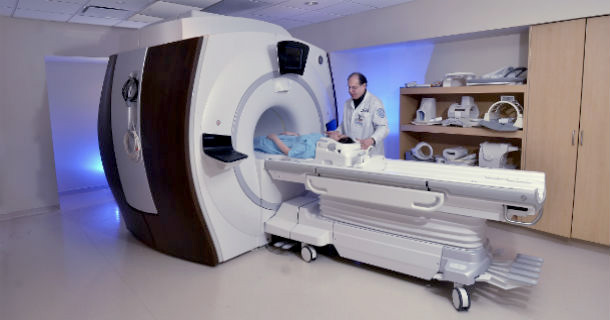
The MRI or MMR MRI equipment has a large ring-shaped magnet and a tunnel inside. This magnet is large enough for the patient to remain inside the tube completely, lying on the stretcher. It also consists of a radio frequency issuer and an antenna that is responsible for receiving the resonances emitted by the body.
The regulation of the intensity of the radio frequencies to which the patient is subjected is carried out by a series of gradient coils. The computer in the technical room is in charge of processing the magnetic signals it receives from inside the cabin and converts them into images.
A patient is usually asked to perform MRI or MRI for both the diagnosis of diseases and pathologies in the bones and in the soft parts of the body. Each inner tissue responds in a completely different way to magnetic radiations. Depending on the area to be defined, the intensity of the magnetic field varies to obtain the cutting-shaped images, in any direction, in two and three dimensions. In this way, the doctor has all the necessary elements for a more accurate diagnosis than with other tests such as CT or ultrasound.
How an MRI is done
The test itself lasts between 25 and 40 minutes, depending on the area to be explored and the organism’s reaction. If the response is insufficient to form clear images, an intravenous contrast is injected.
Its operation is based on radio waves, which are capable of manipulating the magnetic position of matter. This answer is the one that absorbs the antenna so that the computer receives it and turns it into a very high quality image.
Throughout the test, the patient hears a series of noises (more or less intense depending on the test and its duration). Sometimes it may seem like this is a constant hammering. This is caused by the gradients, which are the devices responsible for receiving the body’s response to identify where that signal comes from.
In the face of an MRI, as well as when performing a CT, the patient is required to sign informed consent confirming that his doctor has explained to him what resonance is and what side effects they may feel may be. Throughout the ordeal, you must remain as still as possible. In case of nervousness or inability to control the movement, the patient’s sedation may be done.
When To Do An MRI?
Today, MRI exams are conducted primarily in patients who require a study of the central nervous system, abdominal, and breast area or to be able to diagnose musculoskeletal lesions. The test itself is usually focused on the specific study area to minimize the impact on the rest of the body.
Thus, through MRI, they can be detected in the head from tumors to abscesses and aneurysms that endanger the patient’s health. They are also found through cranial resonance symptoms of internal bleeding, strokes, or nerve lesions originating in the brain. Similarly, they shed light on various pathologies in the eyes and ears, and also of brain degenerative diseases.
In the chest area, MRIs allow specialists to see in a more detailed way the anatomy and state of the heart muscle, as well as the main coronary arteries, in search of some kind of pathology for a more precise diagnosis. Since resonances are suitable for the state of the body’s soft tissues, it has become one of the most important and accurate diagnostic tests for breast cancer and lung cancer.
Similarly, in the pelvic area and abdomen, it is used to see the state of the pancreas, kidneys, or the female and male reproductive system in the face of any signs of tumor, infection, or malformation.
On the other hand, even if their imaging is especially relevant to soft parts, bone diseases can also be detected through an MRI that reveals the existence of arthritis, fractures, ligaments, tendons, or cartilage problems. In this sense, it is widely used for the diagnosis and follow-up of disc hernias.
Finally, it is the baseline test for patients who have iodine allergy. Although magnetic resonance imaging is also used to improve the sharpness of the results, this contrast (gadolinium) does not carry iodine. Hence it is considered more innocuous for the organism.
Cases In Which It Is Contraindicated
Although this is a diagnostic test that can be performed on people of any age, there are people for whom it is not a safe technique. Thus, for example, people suffering from kidney failure may have problems with treatment. Nor can people who have pacemakers or any other implant (audphones, IUDs, or any metal element) do so from it.
People with claustrophobia or very obese who cannot enter the MRI tube.